Prolapsed Gland of the Nictitans (Cherry Eye)
Published on July 14, 2011
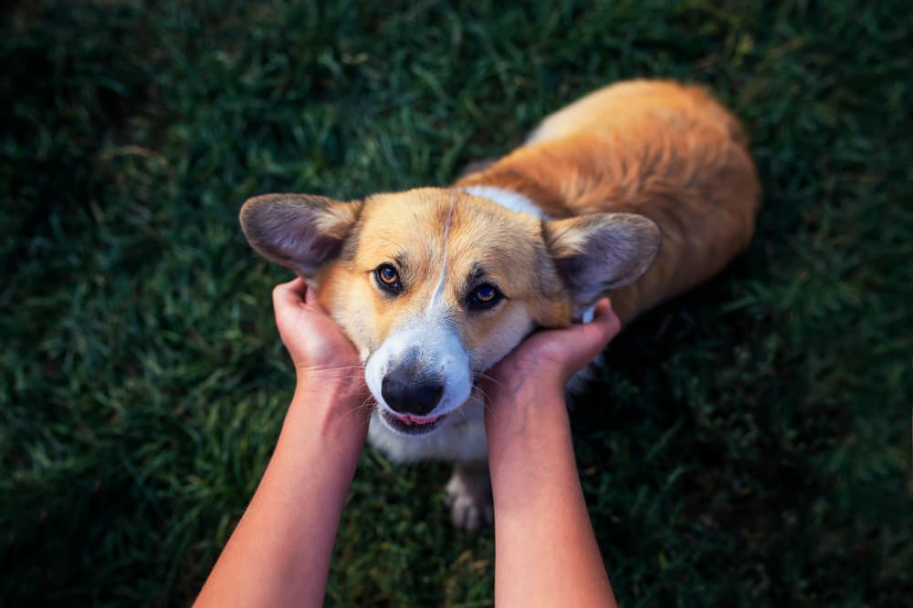
The third eyelid, or the nictitating membrane, is that little triangle in the inner corner of the eye under which a tear gland lives. With some dogs, that gland is displaced, resulting in a condition known as “cherry eye” — so called because the protruding gland become inflamed and sits atop the third eyelid like a rosy fruit. Almost always, surgery is needed to secure the gland back in its natural place.
Overview
The nictitating membrane is a triangular flap of tissue at the inner corner of the eye that’s often referred to as the third eyelid. The entire structure, designed for protection and lubrication of the surface of the eye, includes a flap of cartilage for support and a tear gland located beneath the surface of the lower eyelid.
When the tear gland flips upward (prolapses) out of its normal position under the eyelid and becomes inflamed, the pink, bulbous protrusion that results at the corner of the eye is descriptively referred to as a “cherry eye.”
If untreated, a prolapsed gland of the third eyelid may lead to chronic irritation of the cornea and conjunctiva. In most untreated cases, dry eye (keratoconjunctivitis sicca or KCS) can also result due to decreased tear production. This latter condition can actually progress to severe corneal ulceration and blindness.
Although this ocular condition of canines has not been definitively determined to be hereditary in origin, it’s nonetheless clear that some breeds are predisposed. A complex pattern of inheritance that determines eye conformation is most likely at the genetic root of this problem.
Symptoms and Identification
This condition is very easy to identify, even by non-veterinarians, due to its location and impressive appearance. Dogs will usually experience this prolapse as puppies, most often before 2 years of age. One or both eyes may be affected.
Affected Breeds
The Beagle, Boston Terrier, Bulldog (all breeds), Cocker Spaniel, and Lhasa Apso are all considered predisposed breeds.
Treatment
A prolapsed gland of the third eyelid typically requires surgical treatment. In some very mild cases, a veterinarian or ophthalmologist may successfully replace the gland into its proper position without surgery, but this is atypical. Replacing the gland and surgically securing it in place is almost always necessary. Unfortunately, recurrence of the prolapse will sometimes occur following surgery, necessitating another procedure.
While surgical removal of the gland is still performed by way of treating cherry eyes, this is no longer considered the ideal approach due to the loss of the gland’s tear-producing ability. A high percentage of cases treated this way develop dry eye.
Both general practitioners, veterinary surgeons, and veterinary ophthalmologists can be adept at this procedure. It’s nonetheless important to keep in mind that specialists tend to offer a lower rate of post-surgical recurrence due to their greater experience treating this condition.
Prevention
Based on assumptions as to how this trait may be inherited, all dogs who develop a prolapsed gland of the third eyelid should be spayed and neutered to prevent its propagation.
This article has been reviewed by a Veterinarian.